TLR5 has single Leucine-Rich Repeats (LRR) domain that consists of one N-terminal β-hairpin (LRRNT), 13 complete LRR modules (LRR1-13) and two residues from LRR14.It has two surfaces: the concave surface and the convex surface. The concave surface is formed form 2 anti-parallel β-strands of LRRNT and 13 β-strands of LRR modules. Meanwhile, the convex surface is made up of helices and irregular structures. There are two loops (LRR7 and LRR9) sticking out of the convex surface.

Figure 1.1. The overall structure of TLR5 Zebrafish.
TLR5 has four different types of interface. They are labelled in different colours in figure 1.1. Primary binding interfaces (primary interface-A and B) are coloured green and blue. These two interfaces allow TLR5 to bind to FliC and form a 1:1 heterodimer. The secondary dimerisation interfaces, interface α (cyan) and interface β (red), allow two 1:1 heterodimers to form a 2:2 complex.
TLR5 Disulphide bond
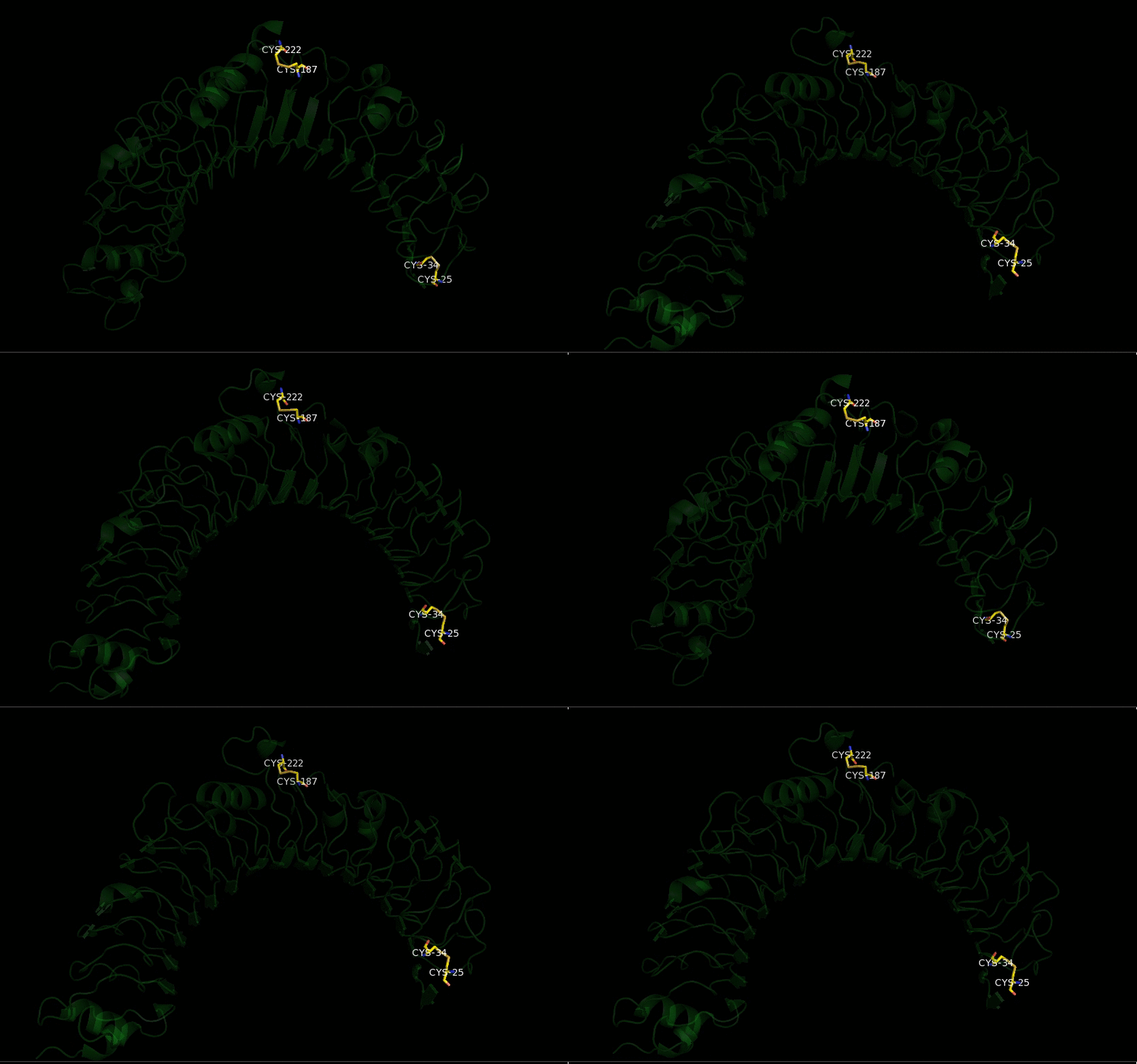
Figure 1.2. 2 disulphide bonds within TLR5.
There are two disulphide bonds within TLR5. Disulphide bond is formed by two cysteine residues. The first disulphide bond is between Cys187 and Cys222 while the second is formed between Cys25 and Cys34.
N-linked glycans in TLR5
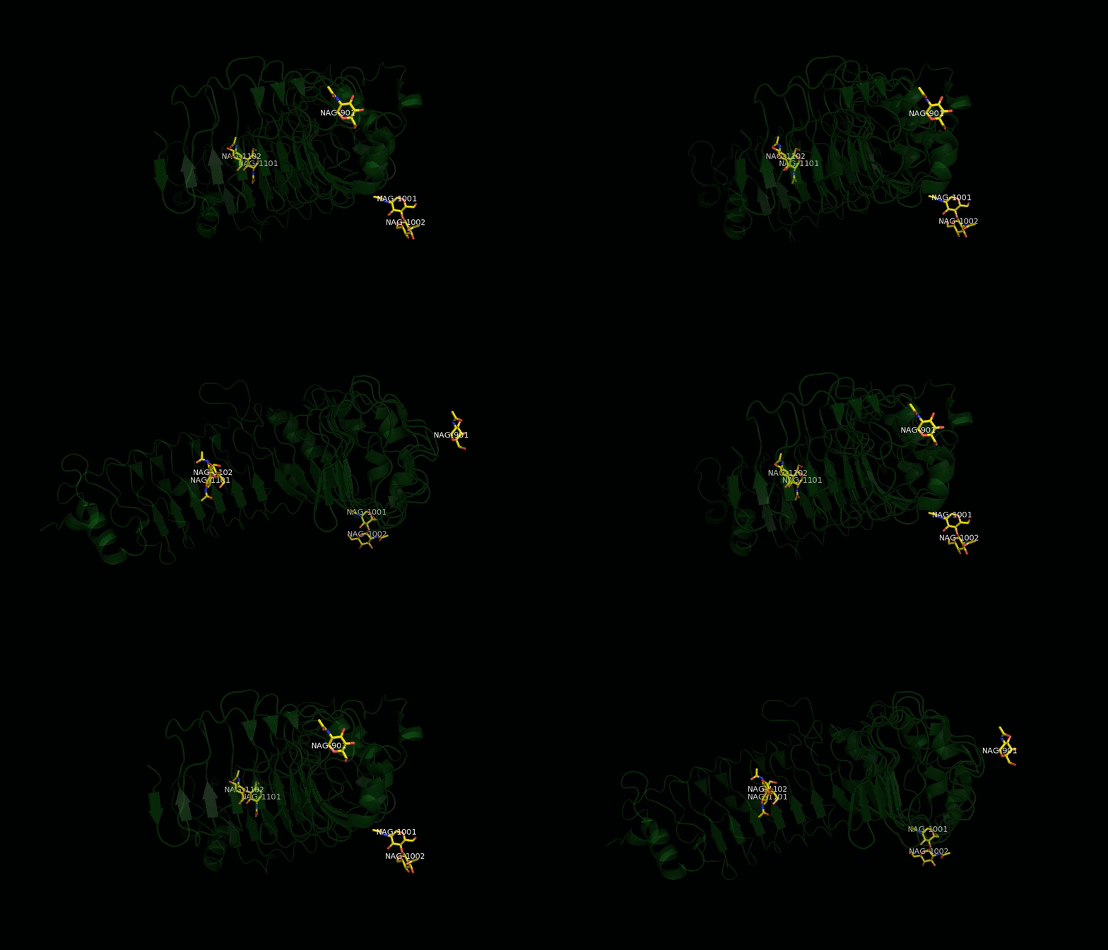
Figure 1.3. 4 N-linked glycans in TLR5.
There are four N-linked glycans in TLR5. These four glycans are scattered over the surface of TLR5. However, they are not involved in any interface.

The GIFs really help to visualise the specific bonds you want to highlight- one comment is to maybe slow down the speed of rotation in the last GIF because it's more difficult to read the labels!
ReplyDeleteThe key at the side of your image is a really good idea because the colouring of each interface is made clear.